
Hemochromatosis: causes,pathophysiology,signs and symptoms,diagnosis and treatment
Hemochromatosis is the abnormal accumulation of iron in parenchymal organs, leading to organ toxicity. It is the most common autosomal recessive genetic disorder and the most common cause of severe iron overload. Signs and symptoms Patients with hereditary hemochromatosis may be asymptomatic (75%) or may present with general and organ-related signs and symptoms. Early symptoms include severe fatigue (74%), impotence (45%), and arthralgia (44%) CLASSIFICATION Hemochromatosis can be classified as Hereditary Hemochromatosis Hemochromatosis, HFE-related (type 1) C282Y homozygosity C282Y/H63D compound heterozygosity Hemochromatosis, non-HFE-related Juvenile hemochromatosis (type 2A) (hemojuvelin mutations) Juvenile hemochromatosis (type 2B) (hepcidin mutation) Mutated transferrin receptor 2, TFR2 (type 3) Mutated ferroportin 1 gene, SLC11A3 (type 4) Acquired Iron Overload,Iron-loading anemias,Thalassemia major Sideroblastic anemia etc Pathophysiology Body-iron content is maintained at 3–4 g such that intestinal mucosal absorption of iron is equal to iron loss. This about 1 mg/d in men and 1.5 mg/d in menstruating women. In hemochromatosis, mucosal absorption is greater than body requirements and amounts to 4 mg/d or more. The progressive accumulation of iron increases plasma iron and transferrin saturation resulting in a progressive increase of plasma ferritin. Since symptoms may be delayed until iron accumulation is excessive, hemochromatosis may not be recognized until total body iron is 10 g, or greater. In women, features are uncommon before menopause because iron loss due to menses ,pregnancy and childbirth tends to offset iron accumulation. Iron overload occurs due to increased iron absorption from the GI tract, leading to chronic deposition of iron in the tissues. Hepcidin is a liver derived peptide which controls basolateral iron transport in the intestine and iron release from macrophages and other cells by binding to ferroportin. Hepcidin, in turn, responds to signals in the liver mediated by HFE, TfR2, and hemojuvelin preventing excessive absorption and storage Iron deposition in the pituitary causes hypogonadotropic hypogonadism in both men and women. Tissue injury results from reactive free hydroxyl radicals generated when iron deposition in tissues catalyzes their formation and disruption of iron-laden lysosomes, from lipid peroxidation of subcellular organelles by excess iron.... Show us your support by SUBSCRIBING,LIKING and SHARING. Connect with us on our website www.medcrine.com Follow us on twitter @medcrine Telegram at https://t.me/medcrine
🎬 More from Medcrine
Anticoagulants: Heparin Mechanisms, Clinical Use, and Monitoring and Clinical Insights
1.2K views
Antidepressants Pharmacology | SSRIs, SNRIs, TCAs, MAOIs, Atypical Antidepressants Simplified
1.0K views
Down syndrome (Trisomy 21) - causes, symptoms, diagnosis, & pathology
376 views
Anencephaly Explained | Neural Tube Defect | Medical and Nursing High-yield Tutorial
1.2K views
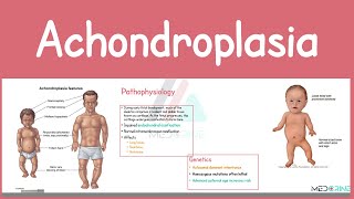
Achondroplasia | Causes, Pathophysiology, Symptoms, Diagnosis, and Management Simplified
959 views
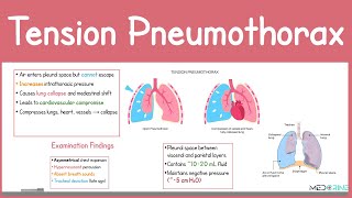
Tension Pneumothorax: Pathophysiology, Clinical signs & Symptoms, Diagnosis and treatment Explained
580 views