
Chest Anatomy on actual CT Scan Simplified with Labels | Radiology Illustration
🛍️ Products Mentioned (1)
Master the anatomy of the thorax on CT using real axial chest CT scan images in this high-yield radiology tutorial. This video walks you through the normal structures of the chest—heart, lungs, vessels, airways, and mediastinum—offering clear explanations ideal for medical and nursing students, radiology trainees, and healthcare professionals. 🔍 In This Video, You Will Learn: Normal chest anatomy on axial CT images Identification of lungs, heart chambers, bronchi, aorta, pulmonary vessels, mediastinum Understanding key anatomical relationships CT interpretation tips for thoracic imaging Become a premium Medcriner TODAY! https://www.youtube.com/channel/UCb1LglhAi_YGQnazlqeWRsQ/join Check our website for more medical lecture notes at https://medcrine.com Follow us on www.x.com/medcrine Facebook at www.fb.me/medcrineacademy Pinterest at pinterest.com/medcrine Telegram at t.me/medcrineacademy email us on info@medcrine.com
🎬 More from Medcrine

Anticoagulants: Heparin Mechanisms, Clinical Use, and Monitoring and Clinical Insights
1.2K views
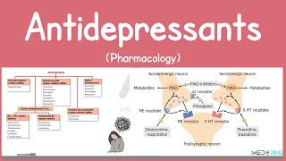
Antidepressants Pharmacology | SSRIs, SNRIs, TCAs, MAOIs, Atypical Antidepressants Simplified
1.0K views
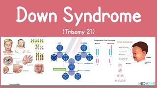
Down syndrome (Trisomy 21) - causes, symptoms, diagnosis, & pathology
376 views
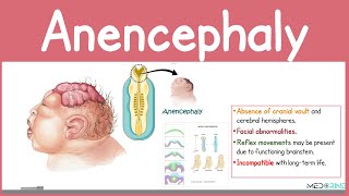
Anencephaly Explained | Neural Tube Defect | Medical and Nursing High-yield Tutorial
1.2K views
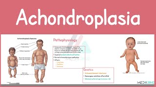
Achondroplasia | Causes, Pathophysiology, Symptoms, Diagnosis, and Management Simplified
959 views
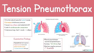
Tension Pneumothorax: Pathophysiology, Clinical signs & Symptoms, Diagnosis and treatment Explained
580 views